Pathophysiology
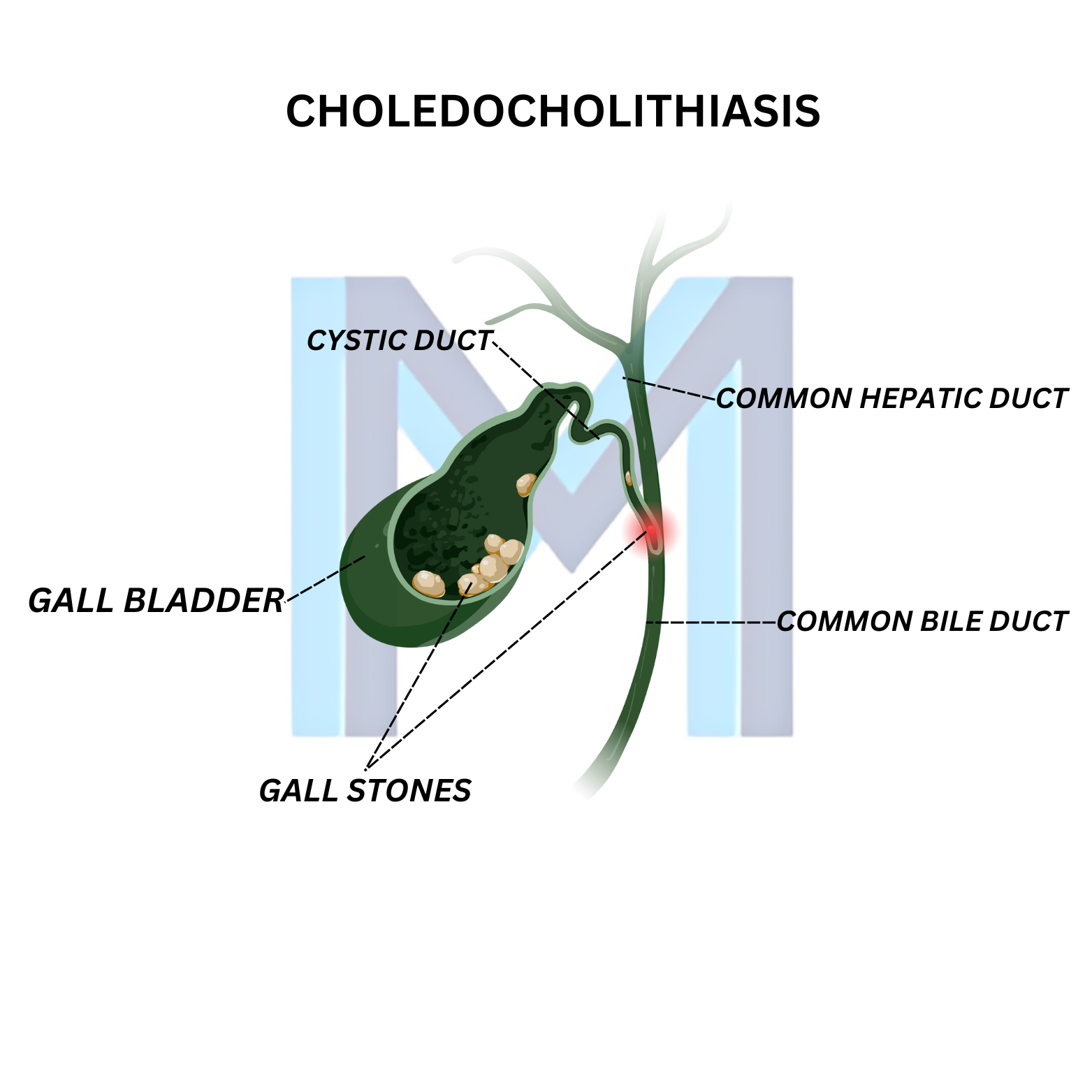
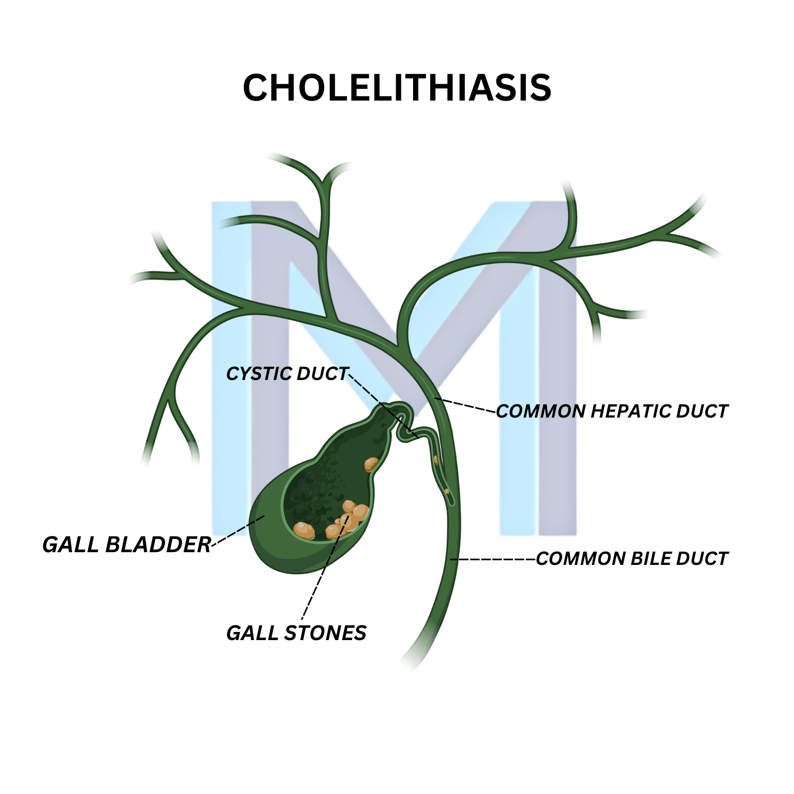
Cholesterol stones (80%): supersaturated bile + gallbladder hypomotility. Pigment stones: chronic haemolysis (black) or infection/stasis (brown). Risk factors classically: female, forty, fertile, fat, family history; also rapid weight loss, TPN, ceftriaxone, ileal disease, sickle cell.
Sources: WSES Guidelines Acute Calculous Cholecystitis (World J Emerg Surg 2020;15:61); Tokyo Guidelines 2018 for Acute Cholangitis/Cholecystitis (J Hepatobiliary Pancreat Sci 2018;25:41)
Clinical presentation
Asymptomatic in ~80%. Biliary colic: RUQ/epigastric pain after fatty meals, lasting 30 min to 6 h, radiating to right scapula, nausea. Fever, jaundice, Murphy sign suggest acute cholecystitis or ductal obstruction.
Sources: WSES Guidelines Acute Calculous Cholecystitis (World J Emerg Surg 2020;15:61); Tokyo Guidelines 2018 for Acute Cholangitis/Cholecystitis (J Hepatobiliary Pancreat Sci 2018;25:41)
Diagnosis
Right-upper-quadrant ultrasound is first-line - stones, wall thickening, pericholecystic fluid, sonographic Murphy sign. HIDA scan for equivocal cases (non-filling gallbladder confirms cholecystitis). LFTs and lipase to detect CBD stone or pancreatitis; MRCP or EUS if choledocholithiasis suspected.
Sources: WSES Guidelines Acute Calculous Cholecystitis (World J Emerg Surg 2020;15:61); Tokyo Guidelines 2018 for Acute Cholangitis/Cholecystitis (J Hepatobiliary Pancreat Sci 2018;25:41)
Management
Asymptomatic stones - observation unless porcelain gallbladder, sickle cell, or bariatric surgery candidate. Symptomatic disease - laparoscopic cholecystectomy. Acute cholecystitis: IV fluids, antibiotics, early laparoscopic cholecystectomy within 7 days. Choledocholithiasis: ERCP with stone extraction, then cholecystectomy.
Sources: WSES Guidelines Acute Calculous Cholecystitis (World J Emerg Surg 2020;15:61); Tokyo Guidelines 2018 for Acute Cholangitis/Cholecystitis (J Hepatobiliary Pancreat Sci 2018;25:41)