Pathophysiology
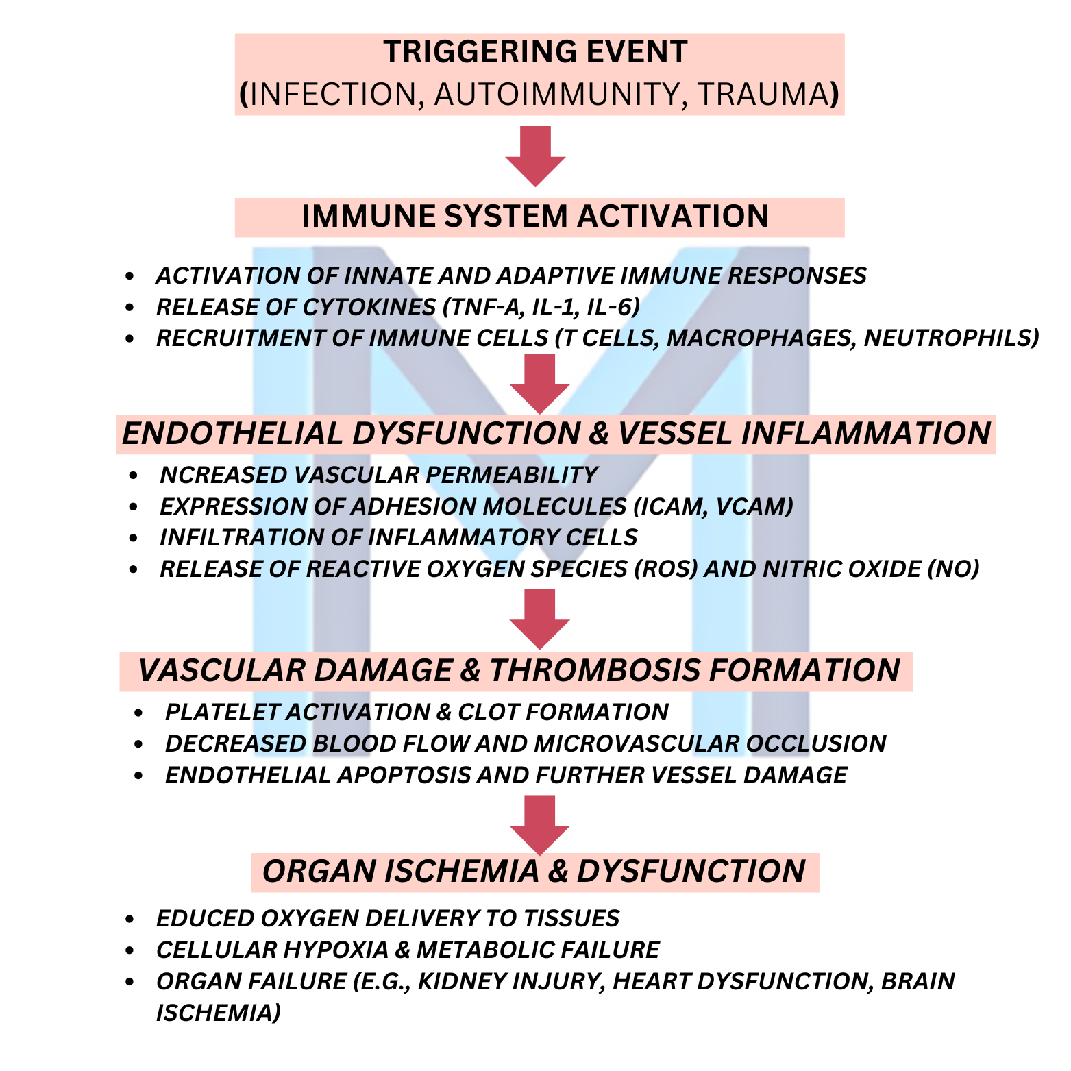
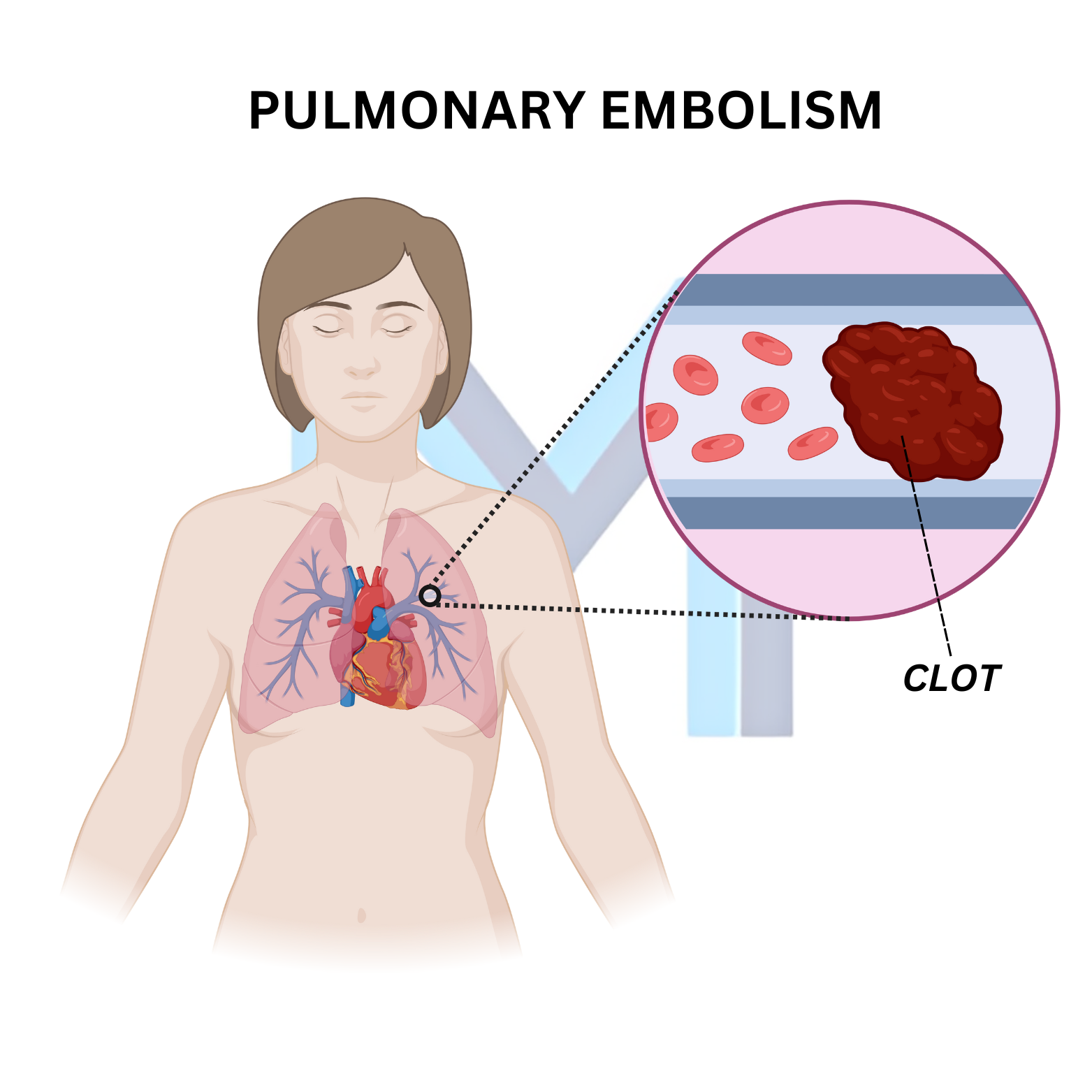
Virchow triad (stasis, hypercoagulability, endothelial injury) predisposes to DVT, most often in the deep veins of the calf and thigh. Embolisation to the pulmonary arteries impairs gas exchange, increases dead-space ventilation and raises pulmonary vascular resistance. Right ventricular afterload rises; massive PE causes RV failure, decreased preload to the LV, obstructive shock and death.
Sources: ESC 2019 PE Guidelines (Eur Heart J 2020;41:543); CHEST 2021 Antithrombotic Therapy for VTE (CHEST 2021;160:e545)
Clinical presentation
Sudden dyspnoea, pleuritic chest pain, cough, haemoptysis, tachycardia, tachypnoea and hypoxaemia. Massive PE: syncope, hypotension, cardiac arrest with PEA. Signs of DVT: unilateral leg swelling, tenderness, warmth. Suspect PE in any postoperative, immobilised, malignancy, pregnancy or hormonal-therapy patient with new dyspnoea.
Sources: ESC 2019 PE Guidelines (Eur Heart J 2020;41:543); CHEST 2021 Antithrombotic Therapy for VTE (CHEST 2021;160:e545)
Risk stratification & diagnosis
Use Wells score. Low probability + negative D-dimer excludes PE. Otherwise CT pulmonary angiography is first-line. V/Q scan is used when CT is contraindicated (renal failure, contrast allergy, pregnancy first-trimester). ECG: sinus tachycardia most common; classic S1Q3T3 is uncommon. Echocardiography assesses RV strain in haemodynamically unstable patients.
Sources: ESC 2019 PE Guidelines (Eur Heart J 2020;41:543); CHEST 2021 Antithrombotic Therapy for VTE (CHEST 2021;160:e545)
Treatment
Haemodynamically stable: therapeutic anticoagulation with a DOAC (apixaban, rivaroxaban), LMWH, or LMWH bridging to warfarin. High-risk (massive) PE with shock: systemic thrombolysis (alteplase) unless contraindicated, or catheter-directed thrombolysis/embolectomy. IVC filter only if anticoagulation is contraindicated or fails.
Sources: ESC 2019 PE Guidelines (Eur Heart J 2020;41:543); CHEST 2021 Antithrombotic Therapy for VTE (CHEST 2021;160:e545)
Duration & prevention
Provoked PE (surgery, trauma): 3 months anticoagulation. Unprovoked or recurrent: consider extended therapy indefinitely, weighing bleeding risk. Cancer-associated PE: DOAC (apixaban, edoxaban) or LMWH for at least 6 months. Prophylaxis: mechanical (IPCs) plus LMWH or DOAC for hospitalised, surgical and postpartum patients.
Sources: ESC 2019 PE Guidelines (Eur Heart J 2020;41:543); CHEST 2021 Antithrombotic Therapy for VTE (CHEST 2021;160:e545)