Pathophysiology
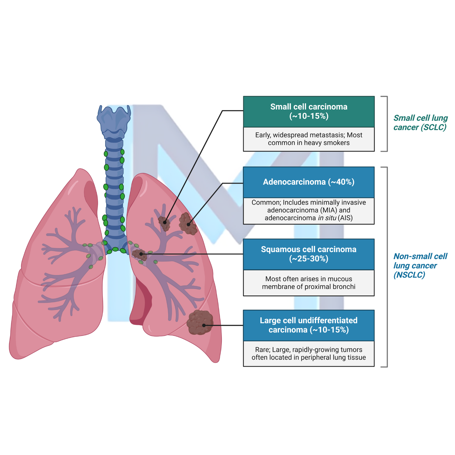
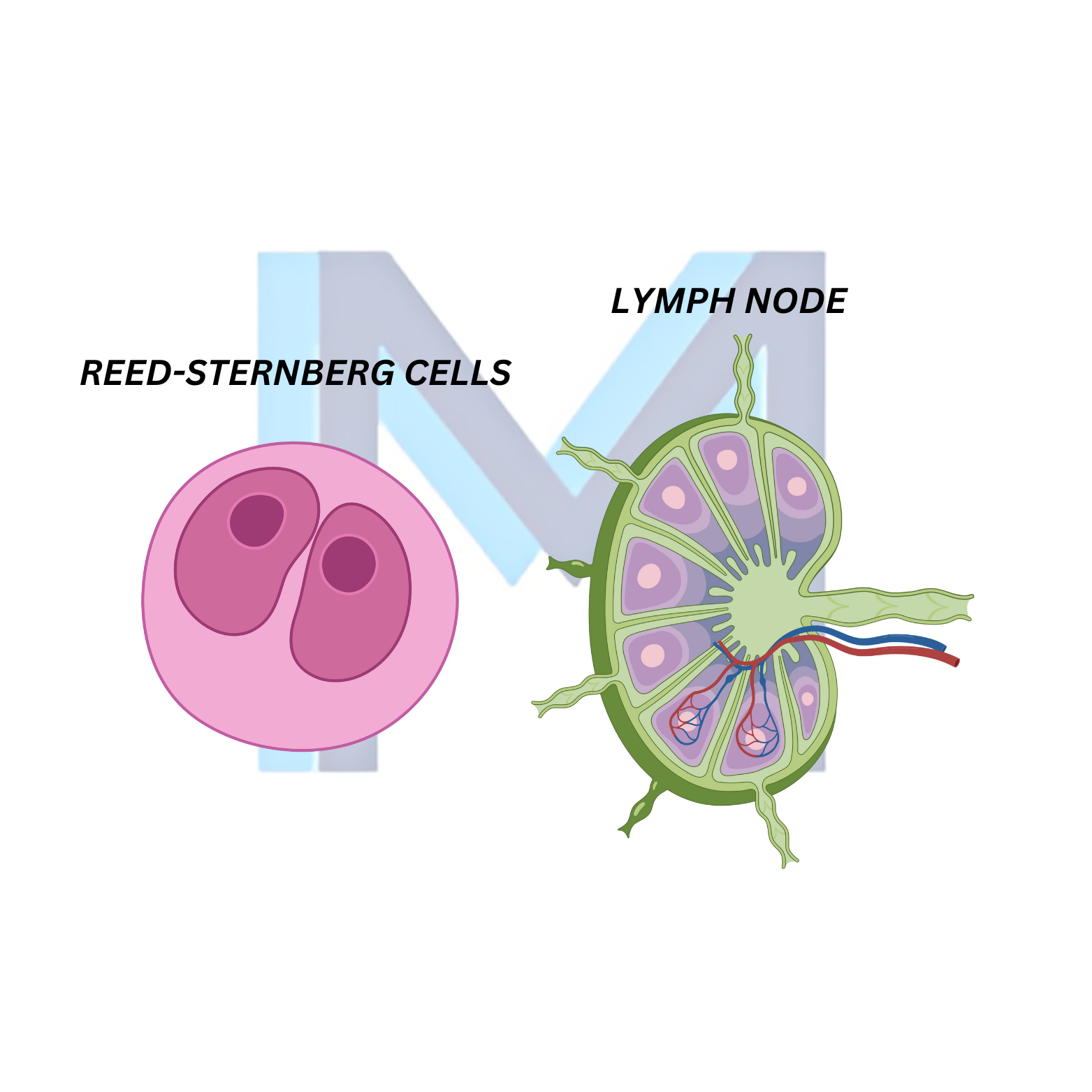
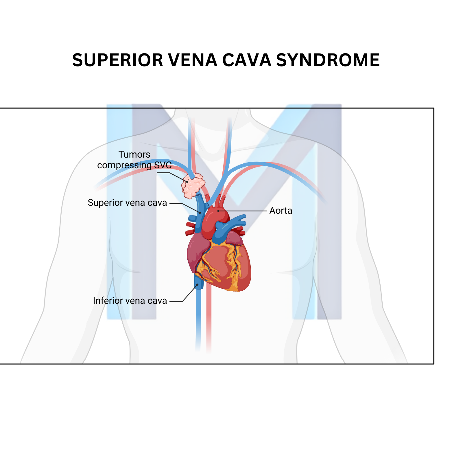
External compression (right-sided lung tumours, mediastinal lymphoma, thymoma) or intraluminal thrombosis (central venous catheters, pacemaker leads) obstruct SVC flow. Collateral flow through azygos and internal mammary systems partially decompresses.
Sources: NCCN Palliative Care - SVC Syndrome (v2.2024); Wilson NEJM 2007;356:1862; ACR Appropriateness Criteria SVC
Clinical presentation
Facial swelling and plethora, distended jugular and chest wall veins, arm swelling, dyspnoea, cough, dysphagia, headache and visual disturbance. Pemberton sign: facial cyanosis on raising arms.
Grading (Yu classification)
Grade 0 asymptomatic to Grade 4 life-threatening (cerebral oedema, laryngeal oedema, cardiovascular collapse) - drives urgency.
Diagnosis
Contrast-enhanced CT chest with venous phase identifies level and cause; MRV if contrast contraindicated. Tissue diagnosis before empiric therapy in stable patients - crucial for lymphoma/germ cell where chemo is definitive.
Management
Elevate head of bed, supplemental oxygen. Grade 3-4: endovascular stent for rapid relief (relief within 24-72 h) followed by treatment of underlying malignancy. Systemic anticoagulation for catheter-related thrombosis, remove catheter when possible. Chemotherapy for SCLC/lymphoma; radiotherapy for NSCLC and radiosensitive tumours; steroids reserved for lymphoma or symptomatic tracheal oedema.