Pathophysiology & types
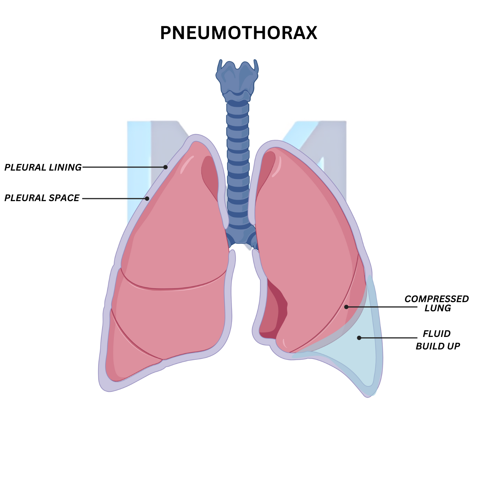
Air enters the pleural space through the visceral pleura (spontaneous/iatrogenic) or the chest wall (traumatic). Loss of negative intrapleural pressure allows the lung to collapse. Primary spontaneous pneumothorax occurs in tall, thin young men from ruptured apical blebs; secondary spontaneous complicates COPD, TB, CF and Pneumocystis. Tension pneumothorax develops when a one-way valve traps air, raising intrathoracic pressure, shifting mediastinum and impeding venous return.
Sources: BTS Pleural Disease Guideline 2023 (Thorax 2023;78:s1); ACCP Consensus statement on pneumothorax management
Clinical presentation
Sudden pleuritic chest pain and dyspnoea. Exam: decreased breath sounds and hyperresonance on the affected side, reduced chest wall movement. Tension: tracheal deviation away, distended neck veins, hypotension, cyanosis - a clinical diagnosis needing immediate action.
Sources: BTS Pleural Disease Guideline 2023 (Thorax 2023;78:s1); ACCP Consensus statement on pneumothorax management
Diagnosis
Erect PA chest X-ray shows visceral pleural line with absent lung markings peripherally. Ultrasound (loss of lung sliding, absent B-lines, lung point) is highly sensitive in trauma. CT is definitive and quantifies size. NEVER wait for imaging in suspected tension pneumothorax.
Sources: BTS Pleural Disease Guideline 2023 (Thorax 2023;78:s1); ACCP Consensus statement on pneumothorax management
Management
Small (<2 cm at hilum) primary spontaneous in stable patient: observation ± high-flow O2. Larger or symptomatic primary: needle aspiration or small-bore chest drain. Secondary spontaneous: chest drain and admission. Tension: immediate needle decompression at the 2nd intercostal space midclavicular line (or 4th-5th intercostal space anterior axillary line in adults per ATLS) followed by chest tube.
Sources: BTS Pleural Disease Guideline 2023 (Thorax 2023;78:s1); ACCP Consensus statement on pneumothorax management
Recurrence & prevention
After a first primary spontaneous, recurrence is 30-50%. Definitive prevention with VATS pleurodesis or bullectomy is offered after second ipsilateral episode, first contralateral episode, persistent air leak, bilateral disease, or in high-risk occupations (pilots, divers). Divers must permanently stop, unless pleurectomy is performed.
Sources: BTS Pleural Disease Guideline 2023 (Thorax 2023;78:s1); ACCP Consensus statement on pneumothorax management