Pathophysiology
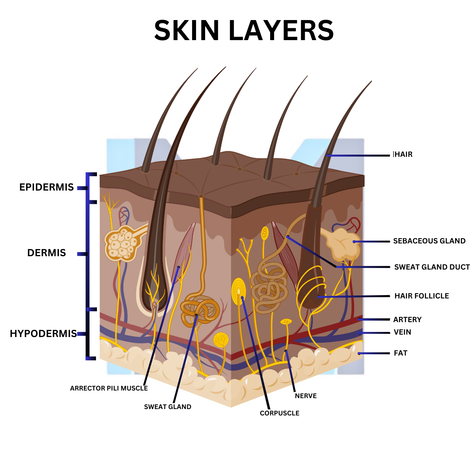
Chronic UV-B exposure induces PTCH1 mutations, activating the Hedgehog/GLI pathway in basal keratinocytes. Fair skin (Fitzpatrick I-II), immunosuppression, arsenic exposure and Gorlin syndrome markedly raise risk.
Sources: NCCN Basal Cell Skin Cancer (v2.2025); AAD Guidelines of Care for BCC (J Am Acad Dermatol 2018;78:540); BAD BCC Guidelines (Br J Dermatol 2021;185:899)
Clinical presentation
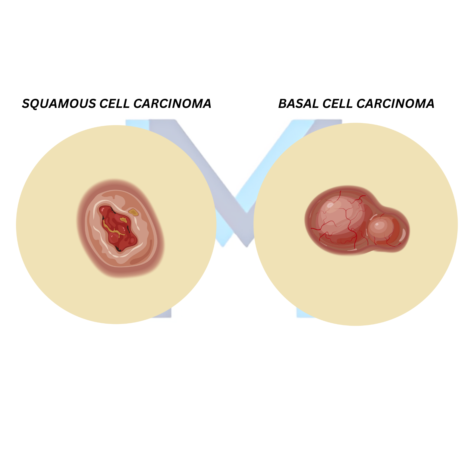
Pearly, translucent papule with rolled borders, central telangiectasia and possible ulceration ('rodent ulcer') on sun-exposed skin (face, ears, scalp). Morpheaform and infiltrative subtypes are scar-like and aggressive.
Diagnosis
Dermoscopy reveals arborising vessels and blue-grey ovoid nests. Confirm with shave or punch biopsy; report subtype (nodular, superficial, infiltrative, morpheaform) which drives treatment.
Management
Low-risk (superficial/nodular, trunk, <2 cm): electrodesiccation and curettage, cryotherapy, topical imiquimod or 5-FU. High-risk or facial 'H-zone': Mohs micrographic surgery. Locally advanced or metastatic: hedgehog inhibitors (vismodegib, sonidegib); cemiplimab for hedgehog-refractory disease.
Prevention & follow-up
Daily broad-spectrum SPF 30+, sun-protective clothing, and full-skin exam every 6-12 months - patients have ~50% risk of a second BCC within 5 years.