The urine dipstick shows 3+ protein and blood. The patient has swollen ankles. Someone says "nephrotic." Someone else says "nephritic." The honest answer, before you have looked at the sediment and measured the protein, is that both may be right.
These terms describe patterns of glomerular injury, not mutually exclusive diseases. Start with what the filter is doing, then name the syndrome 12.
Picture the filter before memorising the list
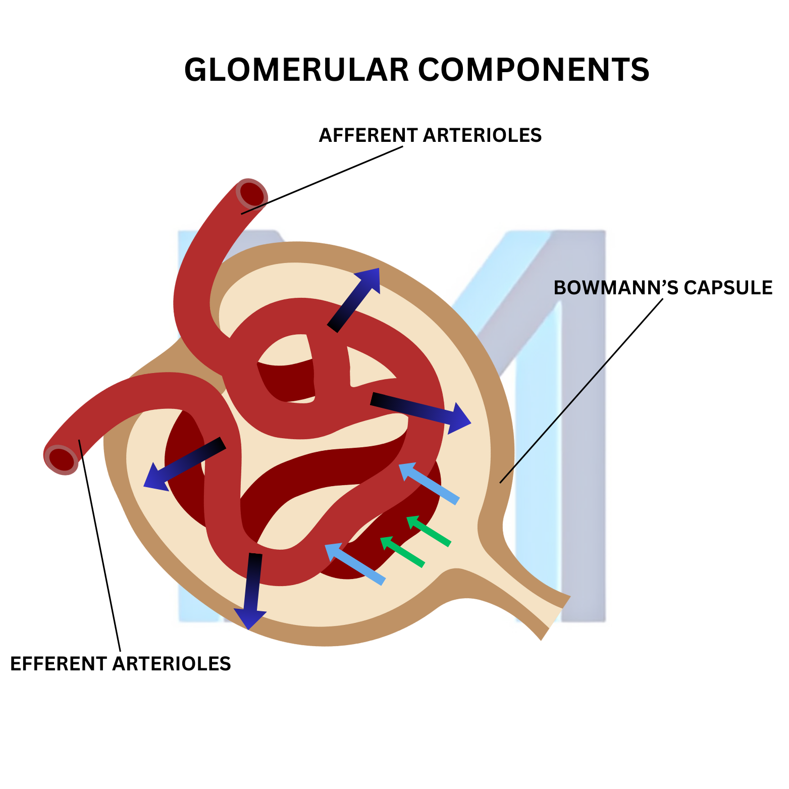
Each glomerulus filters plasma across an endothelial layer, a basement membrane, and podocyte slit diaphragms. If that barrier becomes leaky to protein, albumin escapes into urine. If inflammation injures the capillary tuft, red cells cross too, often alongside a fall in filtration 1.
Nephrotic syndrome is the heavy-protein-loss pattern: marked proteinuria, low serum albumin, and edema, often with hyperlipidemia. Nephritic syndrome is the inflammatory pattern: glomerular hematuria, often red cell casts, reduced kidney function, hypertension, and variable proteinuria 12.
Notice what is not in that description: edema alone. Either syndrome can cause it.
A quick first pass at the bedside
Ask about visible blood or dark urine, recent infections, systemic symptoms, new medicines, and how quickly the swelling appeared. Check blood pressure and volume status. Then use the tests to sharpen, not replace, the story:
- Urine microscopy: dysmorphic red cells or red cell casts support glomerular bleeding. A dipstick positive for blood cannot tell you where the blood came from.
- Quantified protein: use a urine protein-to-creatinine or albumin-to-creatinine ratio rather than treating "3+" as a measured daily loss.
- Serum creatinine and trend: a rapidly rising value changes urgency even when the urine looks only mildly abnormal.
- Serum albumin: marked hypoalbuminemia supports the protein-loss picture, though nutrition and liver disease also affect it 12.
Patterns that are useful, until they aren't
Minimal change disease, membranous nephropathy, and focal segmental glomerulosclerosis commonly enter a nephrotic differential. IgA nephropathy, infection-related glomerulonephritis, and vasculitis often present with a nephritic picture. Lupus can produce either pattern or both 2.
That is why the tidy textbook columns are a starting point rather than a final answer. Heavy proteinuria can accompany inflammatory glomerulonephritis; hematuria can appear in a patient with otherwise nephrotic features. The mechanism and the urgency matter more than forcing a mixed case into one box.
The moment to escalate
New glomerular hematuria with worsening kidney function, marked hypertension, or systemic signs needs prompt assessment. A rapidly progressive picture may require urgent nephrology input and a kidney biopsy to identify the cause before irreversible damage accumulates 2. In a heavy-protein-loss picture, assess the consequences of edema and low albumin, and look for the underlying disease rather than assuming every case needs the same treatment.
Serology and complement testing can narrow the differential when guided by the history and urine findings. Biopsy often establishes the specific diagnosis; the syndrome by itself does not tell you which immunosuppressive drug, if any, a patient needs 2.
The durable mental model is this: a glomerulus can leak protein, bleed, lose filtration, or do all three. Name what you can observe first. The disease name comes after the evidence.