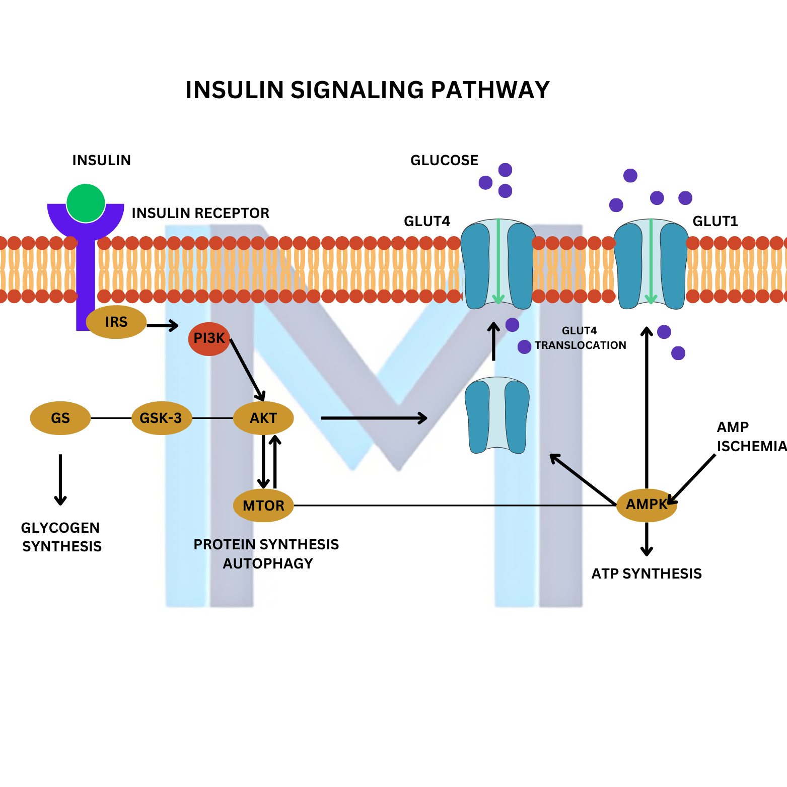
Most students can recite that insulin lowers blood glucose. Far fewer can draw, on a blank page, exactly how that happens inside a muscle cell. That gap is the reason type 2 diabetes pharmacology feels like memorisation. Once the pathway is mapped from receptor to GLUT4, every drug we use snaps into a slot on the diagram.
This is that map. Learn it in the order below and it stays.
The one-line version
Insulin binds a tyrosine kinase receptor, phosphorylates IRS-1, activates PI3K, which activates AKT, which moves GLUT4 vesicles to the plasma membrane and stores glucose as glycogen and fat 1.
Everything in this article is a slower version of that sentence.
1. The insulin receptor is a tyrosine kinase
Two alpha subunits sit outside the cell and bind insulin. Two beta subunits cross the membrane and carry the tyrosine kinase activity on their cytoplasmic tails. When insulin binds, the beta subunits phosphorylate each other on tyrosine residues. Autophosphorylation is the switch. Nothing downstream happens without it.
This is the same architecture as the IGF-1 receptor, which is why very high insulin levels can drive mitogenic effects through the wrong receptor. Keep that in the back of your head when you meet acanthosis nigricans in a severely insulin-resistant patient.
2. IRS-1 is the docking station
The activated receptor recruits and phosphorylates insulin receptor substrate 1 (IRS-1). IRS-1 is not an enzyme. It is a scaffold with phosphotyrosine residues that other proteins latch onto. If you remember one thing about IRS-1, remember this: serine phosphorylation of IRS-1 (rather than tyrosine) shuts the pathway down.
That single detail is the molecular basis of insulin resistance. Inflammatory cytokines from visceral fat - TNF-alpha, IL-6 - and intracellular diacylglycerol from ectopic lipid drive kinases like JNK and PKC-theta that serine-phosphorylate IRS-1, decoupling it from the receptor above and PI3K below 25.
3. PI3K and AKT: the metabolic arm
PI3K (phosphoinositide 3-kinase) binds phosphorylated IRS-1 and converts PIP2 to PIP3 in the inner leaflet of the membrane. PIP3 recruits AKT (also called PKB), where PDK1 and mTORC2 phosphorylate it into its active form.
Active AKT is the workhorse of the metabolic response. Four things to hold on to:
- It phosphorylates AS160, releasing GLUT4-containing vesicles so they can fuse with the plasma membrane.
- It activates glycogen synthesis by inhibiting GSK-3, which unblocks glycogen synthase.
- It stimulates lipogenesis and protein synthesis via SREBP-1c and mTORC1.
- It inhibits hepatic gluconeogenesis by phosphorylating and excluding FOXO1 from the nucleus, shutting down PEPCK and G6Pase transcription.
That last point is why a fasting glucose is such a good readout of hepatic insulin sensitivity. When AKT stops silencing FOXO1 in the liver, the liver keeps pouring out glucose overnight and the fasting number climbs.
4. GLUT4 translocation, in muscle and fat
Muscle and adipose tissue are the insulin-responsive glucose sinks because they express GLUT4, which sits in intracellular vesicles at baseline. Insulin, via the AKT-AS160 step, is what tells those vesicles to fuse with the membrane. No signal, no fusion, no glucose uptake.
Note what is not on that list. The brain, red blood cells and liver rely on GLUT1, GLUT2 and GLUT3, which do not need insulin. That is why hypoglycaemia hits the brain first and why hepatic glucose output is regulated at the level of transcription rather than transporter trafficking 13.
5. The mitogenic arm: Ras, MAPK, and why it matters clinically
IRS-1 also feeds into the Ras/Raf/MEK/ERK cascade, driving proliferation. In healthy physiology this arm is minor. In hyperinsulinaemia it is not. Chronically high insulin selectively drives the mitogenic branch while the metabolic branch fatigues - which is one of the mechanisms invoked to explain the epidemiological link between hyperinsulinaemia, PCOS, and certain cancers 2.
You do not need to memorise the association studies. You do need to be able to say that insulin has two downstream arms and that they can be desensitised independently.
6. How the pathway fails in type 2 diabetes
Insulin resistance is not a single lesion. It is the pathway breaking at three predictable places at once:
- IRS-1 serine phosphorylation from ectopic lipid and inflammatory kinases uncouples the receptor from PI3K.
- PI3K/AKT activity in muscle and adipose falls, GLUT4 stays intracellular, and postprandial glucose rises.
- In the liver, FOXO1 is no longer restrained, gluconeogenesis is not suppressed, and fasting glucose rises.
The pancreatic beta cell compensates by secreting more insulin. That works, for years. When it stops working, fasting glucose crosses the diabetic threshold and the patient walks into your clinic 25.
7. The pathway maps every diabetes drug
This is where the effort pays off. Overlay each class on the diagram and it stops being a list:
- Metformin: reduces hepatic glucose output. It acts largely upstream of the pathway on hepatic mitochondrial respiration and AMPK, restoring FOXO1 restraint indirectly.
- Thiazolidinediones (pioglitazone): PPAR-gamma agonists. They redistribute lipid out of muscle and liver back into subcutaneous fat, lifting the ectopic lipid brake on IRS-1.
- GLP-1 receptor agonists (semaglutide, liraglutide): augment glucose-dependent insulin secretion and suppress glucagon. They are pushing more signal into a partially resistant pathway, and they do it without hypoglycaemia because the effect is glucose-dependent.
- DPP-4 inhibitors: prolong endogenous GLP-1, same logic, smaller effect.
- SGLT2 inhibitors: bypass the pathway entirely by dumping glucose in the urine. Their cardiorenal benefits are the reason the ADA lists them alongside GLP-1 agonists as preferred second-line agents in high-risk patients 4.
- Sulfonylureas: close the beta-cell K-ATP channel, forcing insulin release regardless of glucose. Effective, cheap, and the reason your patient can go hypoglycaemic if they skip a meal.
- Insulin: replaces the missing signal. In type 1 diabetes, it is the entire treatment. In late type 2 it is the honest option.
You did not memorise that list. You read the pathway and predicted it.
8. What to draw before your endocrine exam
On one sheet of paper, from memory:
- Insulin binding the alpha subunits, beta subunits autophosphorylating.
- IRS-1 with two arrows out: PI3K/AKT downwards, Ras/MAPK sideways.
- Under AKT, four boxes: GLUT4 translocation, glycogen synthesis (GSK-3 inhibited), lipogenesis and protein synthesis, FOXO1 exclusion in the liver.
- Three lesion marks on the diagram at IRS-1, PI3K, and the FOXO1 node, labelled insulin resistance.
- Each drug class parked next to the node it targets.
If you can reproduce that sheet in three minutes, you will out-answer most of your cohort on any endocrine question that involves the word insulin.
A closing note
The insulin signaling pathway is the highest-yield diagram in metabolic medicine. It appears in biochemistry, in endocrinology, in pharmacology, and in almost every USMLE Step 1 vignette that opens with obesity or fatigue. The time you spend learning it once, properly, comes back to you for the rest of your training. Draw it tonight. Draw it again tomorrow. Then stop drawing it and start using it.